NeoScience
NEOscience is a company that conducts studies and invents to create the most optimal bioimaging instrument. NEOscience aims at the practicality of bioimaging instrument and the cost-efficiency at the same time. NEOscience’s brilliant ideas and efforts would be served as a foundation for better biological research.
Features
01
REAL COLOR DATA
we use a color camera and optimized filter for the fluorescence signal through the live window without any special analysis. This live window allows you to intuitively identify the position and intensity of the fluorescence and to get image data as it shown.
02
FAST
a fast frame rate capable of recording videos. Due to the fast video speed, many samples can be processed quickly and instantly observed and responded.
03
SIMPLE
a simple, optimized structure, making installation quick and easy. It is also easy to move, manage, and maintain.
04
EASY TO USE
Hardware and software are user-friendly. Filter mounting, exposure control, and image capture are all simple and easy to use.
MULTI FUNCTION
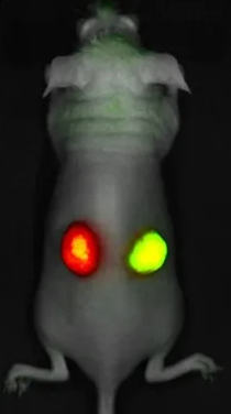
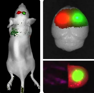
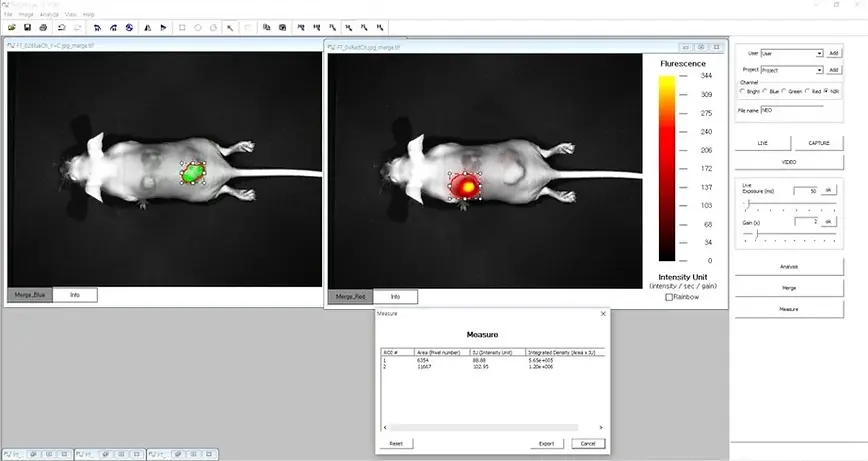
It is possible to apply most fluorescence proteins and fluorescence materials from GFP to ICG using four channels of Blue, Green, Red and NIR. Since more than one fluorescent substance can be imaged, different functions can be observed in one sample. For example, tumor imaging and drug imaging can be performed in the same animal, so targeting and tumorization can be observed simultaneously. You can also merge bright images in order to localization the fluorescence within the animal.
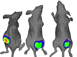
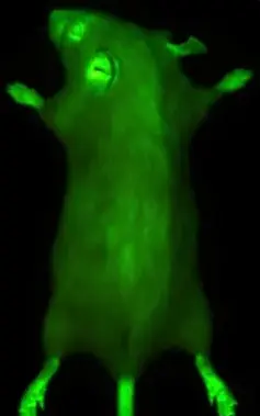
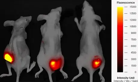
Tumor Imaging
GFP stable cell line can be used to confirm tumorization. The created GFP stable cell line can be imaged In Vitro. GFP cells are injected into subcutaneous tissues and fluorescence images as cell proliferation. In this way, one can obtain images of metastasis to other tissues, in addition to quantifying and comparing tumor size.
Over time, the signal strength of the fluorescence changes, and the camera exposure time may vary accordingly. The NEOimage analysis program can quantify this change by taking into account different conditions such as exposure time and gain; the results of samples with differing images can also be compared and analyzed.
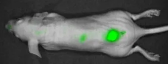
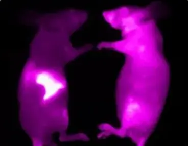
Cell Tracking
Stem cells or immune cells with enhanced functions for various purposes can be imaged within the animal so as to ascertain their location and viability. Stem cells and immune cells are difficult to label with fluorescent genes. So, cells can be stained with fluorescent reagents in a variety of ways.
Stem cells and immune cells stained with a fluorescent reagent can be put into an animal using various methods such as intravenous injection, intraperitoneal injection, and subcutaneous injection.One can determine cell survival using quantitative analysis.
Fluorescence labeled cells can determine the intensity of fluorescence In Vitro. This data can be used to confirm whether or not the fluorescent label is good for In Vivo imaging. This can be used as a basis for predicting or correcting the results of In Vivo experiments. This process can prevent experimental errors. In some cases, the In Vitro experiment can be important in and of itself.
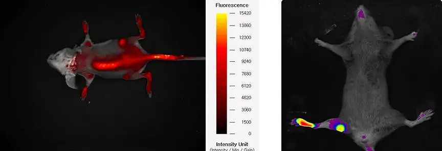
Drugs confirmed In Vitro can be injected into animals for experimental purposes. By taking images at certain intervals, you can check the movement and accumulation pattern of the drug in the living tissues of the animal.
The image of the drug confirmed In Vivo can be checked again Ex Vivo. Because the fluorescence is still expressed even after the animal is sacrificed, it is possible to quantify each tissue separately.
The resulting Ex Vivo data, together with the In Vivo data, can provide excellent evidence for an experiment.

Software - NEOimage
The dedicated software, NEOimage, can capture and analyze fluorescent signals in a very intuitive and easy to use manner. The Live window displays the fluorescent image in real time. It helps determine the optimal exposure time and gain. The fluorescence live window helps you to find the fluorescence signal and observe the operation scene in real time. One can also record a video of the fluorescence signal. All function commands are available as icons in the Dialog bar for easy, intuitive access.
AffiTECHBIO: Revolutionizing Flow Cytometry and Fluorescent Imaging Solutions
At AffiTECHBIO, we are dedicated to empowering researchers and clinicians with advanced tools that elevate the fields of flow cytometry and fluorescent imaging. Our innovative products and solutions provide unmatched precision and flexibility for a wide range of applications in biomedical research, diagnostics, and biotechnology.
Flow Cytometry: Pioneering Excellence with AffiTECHBIO
Flow cytometry is a cornerstone of modern research, enabling high-throughput analysis of cells and particles. AffiTECHBIO supports this critical technology through:
Our portfolio includes a wide range of fluorochrome-conjugated antibodies, tailored for multiplex analyses in flow cytometry.
High specificity and sensitivity reagents for studying complex cell populations.
Precise quantification of rare populations, immune profiling, and functional assays.
Solutions optimized for single-cell analysis, ensuring reproducible and high-fidelity results.
Complement flow cytometry studies by providing robust quantitative assays for protein detection and cytokine analysis, bridging gaps between phenotypic and functional insights.
Why Choose AffiTECHBIO?

Comprehensive Product Range: From flow cytometry reagents to fluorescent imaging tools, we provide end-to-end solutions.

Unmatched Quality: All products are rigorously tested for performance and consistency.

Technical Expertise: Our team of specialists ensures you have the guidance needed to achieve research excellence.

Innovative Kits: Our proprietary AffiELISA® kits integrate seamlessly with flow cytometry and imaging workflows, adding a quantitative dimension to your analyses.